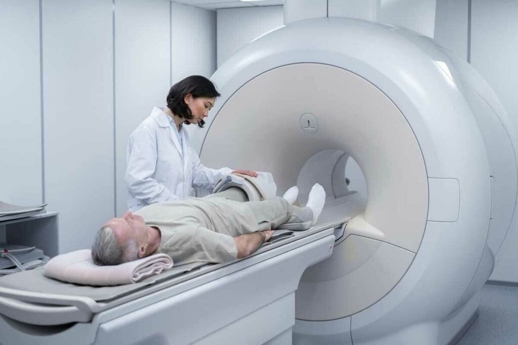
Medical imaging, like X-rays and PET scans, is key for diagnosing and treating diseases. But worries about radiation exposure are on the rise. Every year, about 4.2 billion medical imaging tests are done worldwide, as the United Nations Scientific Committee on the Effects of Atomic Radiation (UNSCEAR) 2020/2021 report shows. It’s important to know the risks these tests pose.
Liv Hospital puts patient safety first. They use strict safety rules. This means every X-ray, mammogram, or PET scan is needed and done with top-notch care.
Key Takeaways
- Medical imaging is essential for diagnosis and treatment.
- Radiation exposure from X-rays and PET scans is a growing concern.
- Liv Hospital adheres to strict safety protocols to minimize radiation exposure.
- Understanding the risks associated with medical imaging is critical for patient safety.
- International-quality care is a priority at Liv Hospital.
The Role of Medical Imaging in Modern Healthcare

Medical imaging has changed how we care for patients. It gives us detailed views of what’s inside the body. If you wonder how many x rays is too many, it is important to know that while there are no fixed limits for medical X-rays for patients, radiation safety guidelines recommend keeping exposure as low as reasonably achievable. The average annual public dose limit from all radiation sources is about 1 millisievert (mSv), and a typical chest X-ray delivers about 0.1 mSv. These guidelines help ensure safe use of imaging in healthcare decisions.
Diagnostic Benefits of Radiation-Based Imaging
Techniques like X-rays and CT scans are vital for finding and treating health issues. They help see bone fractures, tumors, and blood vessel problems.
Key benefits include:
- High-resolution images of internal structures
- Rapid diagnosis, enabling timely treatment
- Guiding minimally invasive procedures
Common Types of Medical Imaging Procedures
There are many medical imaging procedures used today. These include:
| Imaging Modality | Typical Use | Radiation Exposure |
| X-Ray | Bone fractures, lung conditions | Low |
| CT Scan | Detailed internal injuries, tumors | Moderate to High |
| PET Scan | Cancer staging, neurological disorders | Moderate |
Knowing about these imaging methods is important. It helps both doctors and patients make better choices about tests.
Understanding Radiation: Basics and Measurement
Ionizing radiation is used in medical imaging. It’s important to know how it works and how it’s measured. This knowledge helps us understand the risks and benefits of radiation from medical tests.
What Is Ionizing Radiation?
Ionizing radiation has enough energy to remove electrons from atoms. This creates ions. Types include X-rays, gamma rays, and some ultraviolet radiation. In medicine, it helps create images of the body’s inside parts.
The FDA sets rules to keep medical radiation safe. They make sure the benefits of using it for diagnosis are greater than the risks. You can read more about this on the FDA website.
Measuring Radiation: Millisieverts (mSv) Explained
Radiation exposure is measured in millisieverts (mSv). This unit shows the biological effect of radiation. It considers how different tissues react to radiation, giving a more accurate reading.
- Common radiation doses:Chest X-ray: about 0.1 mSv
- CT scan: varies from 2-10 mSv or more, based on the scan type
Effective Dose vs. Absorbed Dose
The absorbed dose shows how much radiation energy is in a tissue mass, in grays (Gy). The effective dose, in sieverts (Sv), looks at the risk of health effects. It considers the sensitivity of different organs and tissues.
An absorbed dose of 1 Gy doesn’t always mean an effective dose of 1 Sv. The effective dose depends on the radiation type and the organs exposed.
Radiation Exposure from Common Medical Procedures
It’s important for patients and doctors to know about radiation doses from medical imaging. These tests help diagnose and treat many health issues. But, it’s key to understand the radiation risks involved.
X-Ray Radiation Levels
A chest X-ray is a common test that uses about 0.1 mSv of radiation. This amount is low, making X-rays safe for many. But, remember, getting many X-rays can add up.
CT Scan Radiation Exposure
CT scans use more radiation than X-rays. They can expose you to 2 to 10 mSv of radiation. This is because they need to show more details for diagnosis.
PET Scan Radiation Amount
PET scans use radioactive tracers and can give a big radiation dose. On average, they expose patients to 8–25 mSv of radiation. The exact dose depends on the procedure and tracer used.
Mammogram Radiation Compared to Other Procedures
Mammograms, for breast cancer screening, use about 0.4 mSv of radiation per view. This is more than a chest X-ray but is considered safe. Comparing it to CT scans and PET scans helps understand the risks better.
Knowing the radiation risks from these tests helps patients and doctors make better choices. This knowledge is key to using these tests wisely.
How Many X-Rays Is Too Many? Addressing the Main Concern
Medical imaging is used more often today. It’s important to know about the risks of getting too much radiation. Getting too many X-rays or imaging tests can harm your health.
Cumulative Radiation Exposure
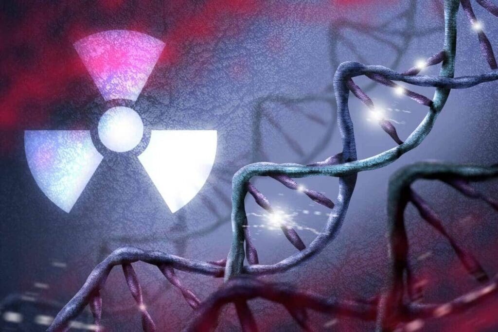
Cumulative radiation exposure is the total radiation a person gets from many tests. The more radiation you get, the higher the risk of health problems like cancer. The body can fix some DNA damage from radiation, but too much can be too much for it to handle.
“The risk of radiation-induced cancer is a function of the total dose received, and the risk is assumed to increase linearly with dose, without a threshold.” – National Research Council
Risk Factors That Influence “Too Many”
Several things can make getting too many X-rays a bigger concern. These include:
- Age: Children are more sensitive to radiation because their bodies are growing.
- Gender: Women are at higher risk for some cancers from radiation.
- Type of imaging: Different tests give out different amounts of radiation.
- Medical condition: Some conditions might need more tests.
Frequency Considerations for Different Imaging Types
Each imaging type has its own radiation level. For example, a chest X-ray has a lower dose than a CT scan. Knowing these differences helps figure out total exposure.
| Imaging Procedure | Typical Effective Dose (mSv) |
| Chest X-ray | 0.1 |
| Mammogram | 0.4 |
| CT Scan (Abdomen) | 10 |
| PET Scan | 8-25 |
Healthcare providers must balance the need for tests against the risk of radiation. They look for the best tests with the lowest dose.
Knowing about radiation risks helps both patients and doctors make better choices. This way, we can use tests wisely and keep health risks low.
Comparing Natural Background Radiation to Medical Imaging
To understand medical imaging radiation, we must compare it to natural background radiation. We are always exposed to small amounts of radiation from our surroundings. This includes cosmic rays from space and radionuclides in the earth, air, and water.
Daily Environmental Radiation Sources
Our daily radiation exposure comes from many sources. These include:
- Cosmic radiation from space
- Radon gas in the air we breathe
- Radiation from the earth’s crust
- Even certain building materials and household items
On average, an individual in the United States receives about 3.1 millisieverts (mSv) of background radiation annually. This amount can change based on where you live, how high you are, and the materials in buildings.
Putting Medical Imaging Radiation in Perspective
Medical imaging procedures like X-rays or PET scans add to our radiation exposure. For example, a chest X-ray gives about 0.1 mSv. A PET scan can give between 8-25 mSv, depending on the type and the tracer used.
To understand the relative risk, consider that a PET scan is equivalent to about 3-8 years of natural background radiation exposure. This comparison helps us see how much radiation we get from medical procedures compared to our daily exposure.
Annual Background Radiation vs. Single Medical Procedures
Let’s compare the annual background radiation dose to the dose from a single medical imaging procedure:
| Radiation Source | Dose (mSv) |
| Annual Background Radiation | 3.1 |
| Chest X-Ray | 0.1 |
| PET Scan | 8-25 |
By comparing these values, we can see that while some medical imaging procedures do increase our radiation exposure, the context of natural background radiation helps in assessing the relative risk.
Radiation Safety Standards and Guidelines
Radiation safety standards are key to protecting patients and workers from harmful radiation. These rules help make sure medical imaging is done safely and well.
Public Exposure Limits
The International Commission on Radiological Protection (ICRP) says the public should not get more than 1 mSv of radiation a year. This limit helps keep the public safe from radiation harm.
Occupational Exposure Limits
People who work with radiation, like radiologists, have a higher limit. The ICRP sets their yearly limit at 50 mSv. They also suggest not going over 100 mSv in five years.
Here’s a table to show the difference between public and worker limits:
| Category | Annual Exposure Limit (mSv) | Five-Year Exposure Limit (mSv) |
| Public | 1 | N/A |
| Occupational | 50 | 100 |
Medical Necessity vs. Radiation Concerns
It’s important to balance medical needs with radiation risks. Doctors must think about the benefits of imaging against the risks of radiation.
Justification is key. Every imaging test with radiation must have a clear medical reason.
Regulatory Bodies and Their Recommendations
Groups like the ICRP and the National Council on Radiation Protection and Measurements (NCRP) give guidelines on safety. They help make rules for protecting against radiation all over the world.
Following these guidelines helps doctors reduce radiation while keeping imaging useful. This way, they can help patients without risking their health.
Understanding PET Scan Radiation in Detail
PET scans are key in modern medicine. They use radioactive tracers to see how the body works. This part talks about PET scan radiation, how they work, and their safety.
How PET Scans Work
PET scans use a radioactive tracer injected into the body. This tracer goes to areas that are very active, like growing cancer cells. The PET scanner then picks up this radiation to show detailed images of the body.
The tracer most often used is Fluorodeoxyglucose (FDG). It’s a glucose molecule with a radioactive atom. Cancer cells, which use more glucose, take up more FDG. This makes them show up clearly on PET scans.
PET-CT Combination and Increased Exposure
Many PET scans are done with CT scans together. This gives more detailed information. But, it also means more radiation for the patient.
A PET-CT scan can give a dose of 10 to 25 millisieverts (mSv). For comparison, a chest X-ray is about 0.1 mSv.
Radioactive Tracers and Their Half-Lives
The tracers in PET scans have short half-lives. This means they lose their radioactivity fast. For example, FDG’s fluorine-18 has a half-life of about 110 minutes.
This short half-life is good because it limits radiation exposure. But, it also means the tracer must be made and given quickly.
Post-Procedure Radiation Considerations
After a PET scan, patients should drink lots of water. This helps get rid of the tracer. The radiation from a PET scan is usually safe. But, patients should avoid being close to others, like kids and pregnant women, for a little while.
It’s smart to talk to your doctor about any worries. They can give advice based on your situation and the scan.
Special Considerations for High-Risk Groups
Some groups face higher risks from medical radiation. This is why special care is needed. Medical imaging is key for diagnosis and treatment, but it must be used wisely in these groups.
Children and Radiation Sensitivity
Children are more at risk because their bodies are growing and they have a longer life ahead. Pediatric patients need lower doses to protect them. Using size-based protocols for CT scans helps reduce their exposure.
| Age Group | Recommended Dose Adjustment |
| 0-5 years | 50% reduction |
| 6-10 years | 25% reduction |
| 11+ years | Standard adult protocol |
Pregnant Women and Fetal Exposure Risks
Pregnant women need special care to protect their unborn babies. The fetus can be harmed by radiation, depending on the dose and when it happens. Alternative imaging methods like ultrasound and MRI are safer choices.
“The use of MRI in pregnancy is considered safe when used appropriately, avoiding the first trimester unless medically necessary.”
Patients with Multiple Medical Conditions
People with many health issues might get more radiation from tests. It’s important to plan each test carefully to avoid too much radiation.
Genetic Factors in Radiation Sensitivity
Genetics play a role in how sensitive someone is to radiation. Certain conditions can make a person more vulnerable. Knowing this can help tailor imaging to each person’s needs.
Healthcare providers can reduce risks by understanding and addressing these special needs. This way, they can protect vulnerable groups from radiation harm.
Discussing Radiation Concerns with Healthcare Providers
When you’re facing medical imaging, talking about radiation with your doctor is key. This chat helps you grasp the risks and benefits. It also helps you make smart choices about your health.
Questions to Ask Before Undergoing Imaging
Before any imaging, ask the right questions. Find out the type of imaging, the radiation dose, and if there are other options. This info helps you understand your options better.
- What type of imaging procedure is recommended for my condition?
- What is the radiation dose associated with this procedure?
- Are there any alternative imaging options that don’t involve radiation?
- How will the results of this imaging procedure impact my treatment plan?
Understanding the Risk-Benefit Analysis
Doctors weigh the benefits against the risks of imaging. They look at your health, the imaging type, and the radiation dose. This helps decide if the imaging is right for you.
| Imaging Procedure | Typical Radiation Dose (mSv) | Benefit |
| Chest X-Ray | 0.1 | Diagnoses lung conditions |
| CT Scan (Abdomen/Pelvis) | 10 | Diagnoses internal injuries or conditions |
| PET Scan | 8-25 | Assesses cancer treatment response |
Requesting Your Medical Imaging History
You have the right to your imaging history. This includes the type and date of past scans. It helps doctors avoid repeating tests and cuts down on radiation.
Tip: Keep a record of your scans. Include the type and date. This helps you and your doctor make better choices.
Advocating for Appropriate Imaging
Patients can push for the right imaging by asking questions and seeking opinions. By working with doctors, you can reduce radiation while getting the care you need.
Alternatives to Radiation-Based Imaging
Concerns about radiation from medical imaging are growing. It’s key to find safer options that don’t use ionizing radiation. Medical imaging has changed a lot, giving us many choices that are safer and effective.
Ultrasound and MRI as Radiation-Free Options
Ultrasound and MRI don’t use ionizing radiation. They are safer for people who need many scans or are sensitive to radiation. Ultrasound uses sound waves to see inside the body. It’s great for looking at organs and checking on babies during pregnancy.
MRI (Magnetic Resonance Imaging) uses a strong magnetic field and radio waves. It’s best for soft tissues like the brain and joints. Both ultrasound and MRI give important info without radiation.
When to Request Alternative Imaging Methods
There are times when to choose non-radiation imaging:
- When non-radiation methods can answer the question.
- For those who have had many scans before.
- In cases where the risks of radiation outweigh the benefits, like for pregnant women or kids.
- When you need to check on something often, using no-radiation methods can lower total radiation.
Emerging Low-Radiation Technologies
New technologies are coming to reduce radiation in medical imaging. Some of these include:
- Low-dose CT scans: New CT scanners use less radiation but keep image quality high.
- Digital X-ray: This uses digital detectors instead of film, lowering doses and speeding up images.
- Advanced image reconstruction techniques: New algorithms help make better images with less radiation.
These new techs are a big step towards safer medical imaging. By using no-radiation and low-radiation methods together, doctors can give better care with less risk.
Conclusion: Making Informed Decisions About Medical Imaging
It’s important to know the risks and benefits of medical imaging. This knowledge helps patients make smart choices about their health. Understanding radiation exposure and safety standards is key.
Radiation safety is a big deal in medical imaging. Knowing the radiation levels of different tests helps patients make better choices. For example, a chest X-ray has a low radiation level, but a PET scan has more.
When deciding on medical imaging, think about the need for the test, its risks, and benefits. Talking to your doctor about your concerns is important. This way, you get the safest and most needed tests.
Being informed and involved in your care is vital. It helps reduce radiation exposure and improves health outcomes. This approach is key for safety and well-being in medical imaging.
FAQ
How much radiation is in a PET scan?
A PET scan can expose you to 8 to 25 millisieverts (mSv) of radiation. This depends on the procedure and the tracer used.
Are chest X-rays dangerous?
Chest X-rays are usually safe, with a low radiation dose of about 0.1 mSv. But, getting many X-rays over time can add up.
How many X-rays is too many?
What’s considered “too many” X-rays varies. It depends on the X-ray type, dose, and your health. Always talk to a doctor about the risks and benefits.
How much radiation do X-rays give off?
The radiation from an X-ray varies. For example, a chest X-ray has about 0.1 mSv. Other X-rays might have more or less.
What is the radiation dose of a mammogram compared to a chest X-ray?
A mammogram has a dose of about 0.4 mSv. This is more than a chest X-ray’s 0.1 mSv. But, it’s also low.
How many X-rays can you have in a year?
There’s no limit to the number of X-rays in a year. It depends on if they’re needed. Doctors try to use less radiation and alternatives when they can.
What are the alternatives to radiation-based imaging?
Ultrasound and MRI are good alternatives because they don’t use ionizing radiation. New, low-radiation technologies are also being developed.
How can I discuss radiation concerns with my healthcare provider?
Ask your doctor about the need for imaging, the dose, and alternatives. Understanding the risks and benefits helps. You can also ask for your imaging history.
Are there special considerations for high-risk groups, such as children and pregnant women?
Yes, children and pregnant women need extra care because they’re more sensitive to radiation. Doctors try to use less radiation and choose safer imaging options.
What are the radiation safety standards and guidelines?
Radiation safety standards include limits for the public (1 mSv annually) and workers (50 mSv annually). The Nuclear Regulatory Commission gives guidelines for safe practices.
How does PET-CT combination affect radiation exposure?
PET-CT combines the radiation from PET scans and CT scans. This means you get more radiation than from a PET scan alone.
What is cumulative radiation exposure?
Cumulative radiation exposure is the total radiation from all imaging procedures over time. It’s key to understanding the risks of repeated exposure.
References
- Smith-Bindman, R., Moghadassi, M., Wilson, N., Nelson, T. R., Boone, J. M., & Cagnon, C. H. (2020). Radiation doses in consecutive CT examinations from five University of California medical centers. JAMA Internal Medicine, *180*(9), 1216-1224. https://www.ncbi.nlm.nih.gov/pmc/articles/PMC7443175/